The intricate connection between the eyes and the brain is facilitated by twelve pairs of cranial nerves. Of these, three are crucial to eye movement: the oculomotor nerve (cranial nerve III), the trochlear nerve (cranial nerve IV), and the abducens nerve (cranial nerve VI). When these nerves become impaired or injured, the result is a condition known as cranial nerve palsy. This condition can cause double vision, misalignment of the eyes, and difficulty controlling eye movement, severely impacting an individual’s quality of life.
 In this post, we will explore the causes, symptoms, and treatment options for cranial nerve palsies that affect the eyes.
What Are Cranial Nerve Palsies?
Cranial nerve palsy occurs when one or more of the cranial nerves responsible for eye movement become damaged or dysfunctional. This can result in weakness or paralysis of the muscles they control, leading to abnormal eye positioning, strabismus (eye misalignment), or diplopia (double vision).
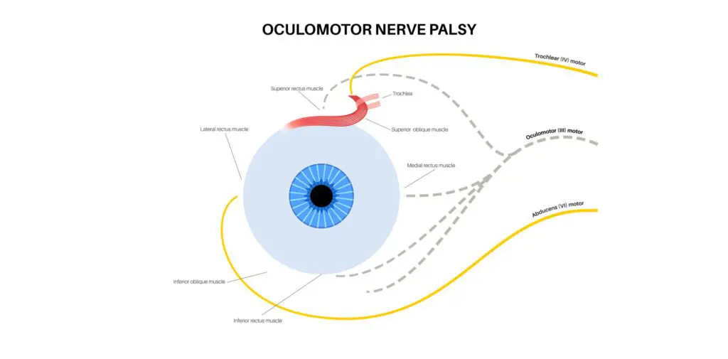
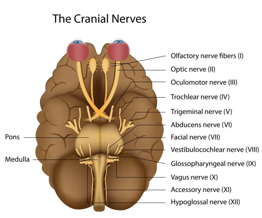 The three cranial nerves that control eye movements are:
1. Oculomotor nerve (CN III): Controls most of the eye’s movements, including those of the eyelids and the muscles that constrict the pupils.
2. Trochlear nerve (CN IV): Controls the superior oblique muscle, which allows the eye to move downward and inward.
3. Abducens nerve (CN VI): Controls the lateral rectus muscle, which allows the eye to move outward (laterally).
Damage to any of these nerves can interfere with the affected individual’s ability to control eye movements, leading to a range of symptoms.
Causes of Cranial Nerve Palsies
The underlying causes of cranial nerve palsy can vary widely, and understanding these causes is critical to determining the appropriate treatment plan. Here are some of the most common causes:
1. Trauma:
Head injuries can result in cranial nerve palsies, especially when there is direct trauma to the nerve pathways that control eye movements. Car accidents, falls, or sports injuries are common traumatic causes.
2. Stroke:
A stroke can disrupt blood flow to the cranial nerves, causing nerve damage that affects eye movement. Strokes affecting the brainstem are particularly likely to involve cranial nerves III, IV, or VI.
3. Aneurysms:
An aneurysm, which is a ballooning of a blood vessel, can compress one of the cranial nerves. An aneurysm near the oculomotor nerve, for example, can cause oculomotor nerve palsy, leading to eyelid drooping (ptosis) and double vision.
4. Infections:
Certain infections, such as meningitis or Lyme disease, can lead to inflammation of the cranial nerves, disrupting their ability to function properly.
5. Diabetes:
Diabetes can cause a condition known as microvascular cranial nerve palsy. Elevated blood sugar levels damage the small blood vessels that supply the cranial nerves, leading to nerve impairment. This condition is more common in older adults and may affect the oculomotor or abducens nerves.
6. Tumors:
Tumors in the brain or surrounding structures can press on the cranial nerves and interfere with their function. Both benign and malignant tumors can lead to cranial nerve palsy.
7. Idiopathic Causes:
In some cases, the exact cause of cranial nerve palsy is unknown, and it is classified as idiopathic. While these cases may resolve on their own, they can still present with significant symptoms that require management.
Symptoms of Cranial Nerve Palsies
The symptoms of cranial nerve palsy can vary depending on which nerve is affected. However, common symptoms include:
1. Double Vision (Diplopia):
One of the hallmark symptoms of cranial nerve palsy is double vision. This occurs because the eyes are no longer aligned properly and are sending two different images to the brain.
2. Eye Misalignment (Strabismus):
The affected eye may turn in, out, up, or down depending on which nerve is involved. For instance, damage to the abducens nerve often results in the eye turning inward.
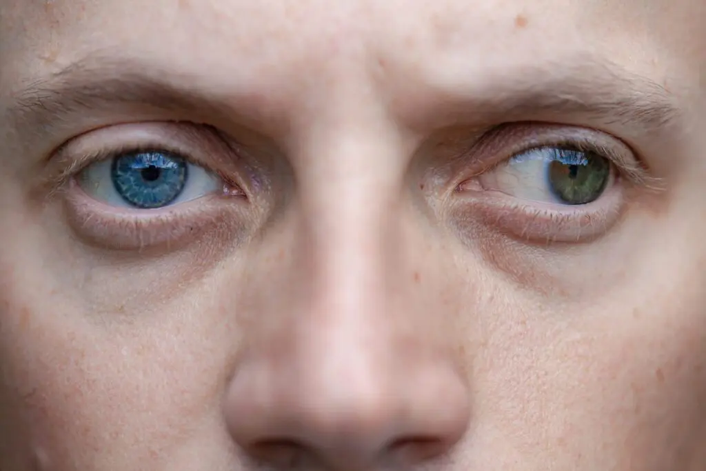
3. Difficulty Moving the Eye:
Affected individuals may notice that they have trouble moving their eye in certain directions. For example, an oculomotor nerve palsy may prevent upward or inward movement, while a trochlear nerve palsy may make it difficult to look downwards.
4. Ptosis (Drooping Eyelid):
Oculomotor nerve palsy often causes ptosis, where the upper eyelid droops because the muscle controlling it is weakened.
5. Pupil Abnormalities:
The oculomotor nerve also controls the muscles responsible for pupil constriction, so individuals with oculomotor palsy may notice that one of their pupils is larger or slower to respond to light.
6. Head Tilt or Turn:
Some individuals develop a compensatory head tilt to reduce double vision. For instance, people with trochlear nerve palsy may tilt their head to one side to help align their vision.
Diagnosing Cranial Nerve Palsies
Diagnosis of cranial nerve palsy begins with a detailed history and clinical examination. An eye care professional will carefully assess the eye’s movements, pupil responses, and eyelid function. They will also ask questions about the onset of symptoms and any possible risk factors, such as recent trauma or medical conditions.
Additional tests may include:
-
Imaging (MRI or CT Scan): Imaging tests are crucial for identifying any structural abnormalities, such as tumors or aneurysms, that may be compressing the cranial nerves.
-
Blood Tests: Blood tests can help identify underlying conditions, such as diabetes or infections, that may contribute to cranial nerve palsy.
-
Neurological Examination: A full neurological examination may be necessary to determine if other cranial nerves or parts of the nervous system are involved.
Treatment Options for Cranial Nerve Palsies
The treatment for cranial nerve palsy depends largely on the underlying cause. In many cases, addressing the root of the problem will lead to improvement in symptoms. Treatment options include:
1. Observation:
Some cases of cranial nerve palsy, particularly those caused by minor trauma or microvascular issues (like diabetes), may resolve on their own with time. Observation and regular follow-up appointments are often recommended in these cases.
2. Prism Glasses:
Prism lenses can help manage double vision by realigning the images seen by the eyes, reducing strain and improving visual clarity.
3. Eye Patching:
In cases of severe double vision, patching one eye may temporarily alleviate symptoms while waiting for the condition to resolve or for other treatments to take effect.
4. Vision Therapy:
Vision therapy can be a helpful non-surgical approach for retraining the brain and eye muscles to work together more effectively. Exercises can help improve eye alignment and reduce symptoms like double vision.
5. Surgery:
In some cases, surgery may be necessary to correct misalignment of the eyes, particularly if the condition is not resolving on its own or if the misalignment is severe. Surgical options may include muscle tightening or repositioning to improve eye movement.
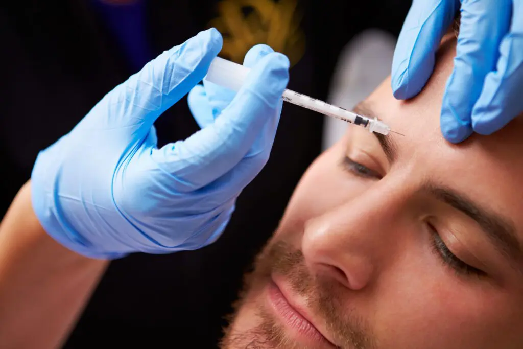
6. Botox Injections:
Botox can be used to weaken overactive muscles, helping to improve alignment and reduce double vision in some patients with cranial nerve palsies.
7. Treating the Underlying Condition:
If the cranial nerve palsy is caused by a systemic issue, such as diabetes or a tumor, treating the underlying cause is critical. Managing blood sugar levels or surgically removing a tumor may result in significant improvement of symptoms.
Cranial nerve palsies affecting the eyes can disrupt daily life with discomfort and vision problems. However, with a proper diagnosis and the right treatment plan, many people see substantial improvement. Treatments like prism glasses, vision therapy, or surgery can help manage symptoms, while addressing the underlying cause is crucial for restoring eye function and enhancing overall quality of life.
Until next month,
Dr. Paul Rollett, OD, FCVOD

